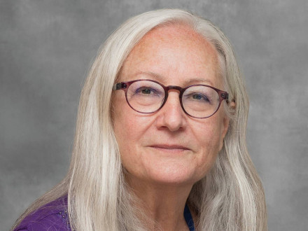
Bruno Passebon Soares, MD, is an Associate Professor of Radiology at Stanford University School of Medicine and serves as the Section Chief of Pediatric Neuroradiology at Lucile Packard Children’s Hospital at Stanford since June 2023. Born in Brazil, Dr. Bruno P. Soares obtained his medical degree from the Federal University of Rio de Janeiro and completed his residency in Diagnostic Radiology at the Federal University of Sao Paulo, followed by fellowship training at the University of California San Francisco. His research has focused on neonatal and pediatric brain imaging.
You deal with the issue of abusive head trauma in children, which involves diagnostics that can eventually lead to charges etc. I know, even from personal experience, that there are cases when individual radiologists’ views can differ strikingly. How reliable is radio diagnostics for head traumas and how to ensure errors will not happen?
This is an important and often controversial topic. Radiologic interpretation is inherently a process of pattern recognition and probabilistic assessment. While certain findings can raise suspicion for abusive trauma, no single imaging finding is pathognomonic in isolation. The crucial factor when interpreting diagnostic imaging exams of suspected abusive head trauma, in addition to deep knowledge of injury mechanisms and correct terminology, is to understand and integrate the clinical context. Often, the imaging findings in isolation are not diagnostic of abusive trauma. In my personal experience, the interpretation of findings performed by experienced and intellectually honest radiologists are largely concordant. Quoting Sir William Osler, “the practice of medicine is an art based in science” and we should strive to adhere to current professional guidelines. Finally, these cases should always be evaluated in conjunction with a multidisciplinary team. At Stanford, we are fortunate to work with the SCAN – Suspected Child Abuse and Neglect – team which includes physicians – pediatricians, radiologists, surgeons –, social workers, psychologists, nurse practitioners, and child life specialists. The team provides comprehensive medical evaluations, offers mental health services for children and families, and works closely with law enforcement and child protective services.
What is your recent success in development of algorithms for detecting cerebral abnormalities in children with epilepsy?
Computer-aided detection of cerebral abnormalities on brain MRI exams of children with epilepsy is a challenging and exciting area of ongoing work by our group at Stanford and elsewhere. Our recent focus involves leveraging deep learning and machine learning to better detect and characterize subtle epileptogenic lesions occasionally missed by visual analysis. We are creating a machine learning algorithm and a radiology-defined MRI database. Our next step is to train this algorithm using brain MRI foundation models and then test its performance on our database. Longer-term, we plan to create our own foundation model with thousands of pediatric brain MRI exams, which we anticipate will significantly improve the algorithm's performance. In addition, we believe multimodal information will improve the quality of the automated analysis, therefore we plan to integrate PET imaging, EEG findings, and clinical and pathological findings whenever available.
Sight plays a key role in image evaluation, and not just of the content of the image but also its quality. Your friends think you are a wine connoisseur. Are you demanding of visual quality also in wine or food? What do you appreciate about the colour and clarity of wine?
I have wonderful friends who may be playfully exaggerating my abilities. My wine tasting expertise and vocabulary reflect much more enthusiasm than proficiency! In fact, I probably read more about wine regions than taste all their wines. But as an example, the color of a wine can hint at the grape varietal, its age, and even the region it hails from. A deep ruby red might suggest a young wine, while a brick-red tinge could indicate a more mature vintage. When thinking about clarity, it may indicate winemaking process, for example the extent of filtration, and also the age of a wine. While brilliant clarity in wine is visually appealing, the presence of sediment can be a natural byproduct and hint at a wine's age, minimal intervention, and potential complexity. Both clear and slightly cloudy wines can be appreciated for what their visual presentation suggests about their journey.
Do you prefer vision over hearing in the arts, e.g., painting over opera?
While I enjoy visual arts, particularly classic paintings, I am much more a history and culture aficionado. In preparation for the visit to Prague, I borrowed several books at the Stanford libraries. Most recently, I have enjoyed reading The Golden Maze by Richard Fiddler. From Good King Wenceslas to the Good Soldier Švejk (also a book title) and beyond, I am finding the history of the Czech lands and its peoples a fascinating topic!
Související
Motol Day of Imaging in Paediatric Radiology / Masterclass
29 May 2025
9:00—17:00
Great Lecture Hall, Motol University Hospital
For Me, Teaching and Learning Is a Two Way Street, Says Stanford Professor Beverly Newman
At the upcoming Motol Day of Imaging in Paediatric Radiology / Masterclass, she will deliver a lecture titled Late Presentation, Complications, and Mimicry of Congenital Pulmonary Anomalies. A conversation.
I Love the Collaborative Environment of Children’s Hospitals, Says Dr Jesse Kerr Sandberg
A conversation with a Stanford radiologist and speaker at the Motol Day of Imaging in Paediatric Radiology, scheduled for 29 May 2025.
Contrast Enhanced Ultrasound Provides Additional Information to Regular Ultrasound About Perfusion of Organs
A conversation with a Stanford radiologist and one of the speakers at the Motol Day of Imaging in Paediatric Radiology, scheduled for 29 May 2025.
If You Don’t Like Your Place/Specialty/Life, Make a Change, Don’t Complain
A conversation with a Stanford radiologist and one of the speakers at the Motol Day of Imaging in Paediatric Radiology, scheduled for 29 May 2025.
Doctors Care About the Safety of the Child Patient, but Parents Should Not Feel Blamed When Communicating With Them
A conversation with Dr Eliška Popelová, a speaker at the Motol Day of Imaging in Pediatric Radiology (29 May 2025).


